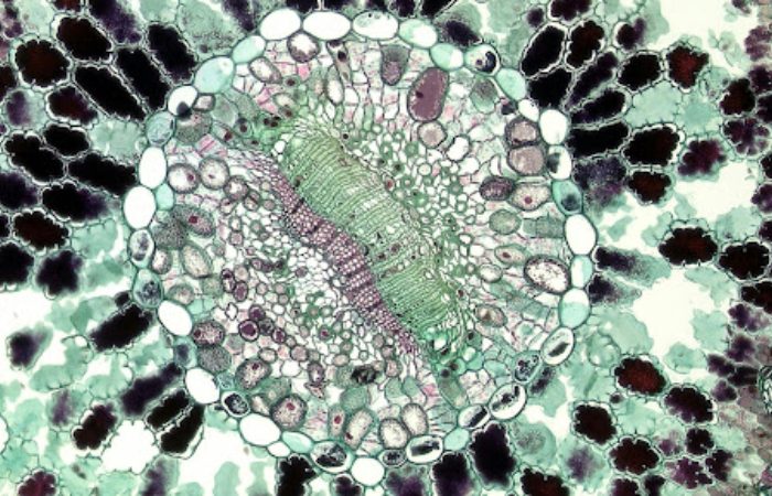
Peering into the microscopic world is like flipping on a light in a dark room—what’s hidden suddenly snaps into view. Fluorescent microscopy takes that a step further, turning tiny, invisible wonders into glowing masterpieces. It’s not just cold science; it’s a dance of light and color, blending precision with a painter’s flair.
Scientists use it to spy on cells, proteins, and life’s smallest gears, revealing secrets that shape medicine, biology, and beyond. From a lab bench to a gallery wall, this tool’s magic lies in making the unseen not just visible, but breathtaking—a fusion of art and discovery that keeps us staring, wide-eyed.
Lighting Up the Tiny World
Picture a cell under a lens, dull and gray—then zap it with a burst of light, and it flares into neon life. That’s the heart of fluorescent microscope images: dyes or proteins that glow when hit with specific wavelengths. Think of a scientist tagging a neuron with a green marker—it’s dark until the scope’s beam strikes, and suddenly, that neuron’s a vivid streak against a black void.
It’s not random; those tags latch onto exact targets—DNA, mitochondria, whatever’s in the crosshairs—turning a blob into a map. The result’s a snapshot, sharp and surreal, where every glow tells a story of what’s ticking inside.
The Science Behind the Shine
It’s not magic—it’s physics with a twist. Fluorescent molecules soak up light energy, get excited, and spit it back out at a different wavelength—think absorbing blue and beaming green. The microscope’s the maestro, wielding filters and beams to catch that shift.
A laser might ping the sample, a lens grabs the glow, and a detector paints the picture. It’s picky—only the tagged bits shine, so the noise fades, and the target pops. That clarity’s why it’s gold for peering into cells—dissecting a cancer quirk or tracking a virus’s sneak attack, all in living color.
Artistry in the Lens
But it’s not just data—there’s a brushstroke vibe here. Those glowing reds, blues, and yellows could hang in a gallery, and sometimes they do. Scientists tweak the hues—pairing a protein with a dye that fits the mood or the mission. A nerve cell might shimmer gold, a nucleus pulse purple—it’s functional, sure, but it’s also a choice, a splash of style.
Zoom in, and it’s a cosmic swirl; pull back, and it’s a living abstract. That beauty’s no accident—it’s a hook, drawing eyes to the science and making the invisible feel real, even poetic.
Peeking at Life’s Playbook
This isn’t just for show—it’s a glimpse into the inner workings of life. By tagging a protein with fluorescence, you can track its movement—gliding along the cell’s framework or gathering with its team. It’s like tailing a suspect: where’s it going, who’s it meeting? That’s clutch for cracking how diseases tick—say, spotting where Alzheimer’s tangles clog or how a flu bug invades. The microscope catches it live—no freeze-frame guesses—just raw, glowing action. It’s a backstage pass to biology’s drama, lit up for all to see.
Pushing the Edges
The tech’s not static—it’s a restless tinkerer’s dream. Super-resolution tricks now zoom past old limits, shrinking the blur to pin down molecules in tight knots. Multi-color tagging lets you track a whole posse—red for this, green for that—all at once.
Time-lapse flips it into a movie: cells splitting, glowing, shifting over hours. It’s not cheap—fancy scopes and dyes cost a mint—but each leap sharpens the view, turning fuzzy blobs into crisp portraits. The invisible’s not just lit; it’s in high-def now.
Hands-On Craft
There’s a craft to it too—part lab coat, part artist’s smock. Prepping a sample’s a ritual: pick the right dye, coax it into the cell, tweak the light so it sings. Too much glow, and it’s a washout; too little, and you’re squinting at shadows.
Scientists play with settings—brightness, focus, filters—like dialing in a perfect shot. It’s trial and error, a feel thing—knowing when the image pops just right. That hands-on grind’s what turns raw science into those jaw-dropping fluorescent microscope images you can’t unsee.

Final Thoughts
This mashup of art and science doesn’t just stay in labs—it spills out. Museums showcase these shots—cells like fireworks, neurons like constellations—hooking folks who’d never crack a textbook. It’s a bridge: researchers geek out on the how, while the rest of us marvel at the wow. Teachers lean on it too—nothing sells a kid on biology like a glowing squid cell. It’s not about dumbing down; it’s about lighting up—making the tiny feel huge, the complex feel close. That’s the real glow: a spark that sticks, from the scope to the soul.


